other:dsx:x4d:x-ray_and_drr_settings
Table of Contents
X-Ray and DRR Settings
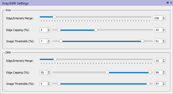
The X-Ray and DRR Settings widget allows users to specify the parameters to use when matching digitally reconstructed radiograph (DRR) images with X-Ray images. This process is sensitive to the image processing parameters in this widget. Once the DRR images have been generated for a particular set of bone poses, the DRR images and the X-ray images are processed (using an identical method), and then compared to each other using the selected image metric algorithm.
Processing Method
At a high level the processing method consists of:
- performing a Sobel edge detection on the image,
- thresholding the edge-detection image
- multiplying the edge-detection image by a weighting factor and adding it to the original image, and
- thresholding the merged image.
You can learn more about considerations for this matching here.
Parameters
| Parameter | Description |
|---|---|
| Edge/Intensity Merge | This is the value of the weighting factor in step 3. |
| Edge Capping (%) | These two values values govern the edge thresholding in step 2. Pixels in the edge image whose values are greater than the edge capping maximum are set to zero. Very bright pixels (sharp edges) are usually inorganic objects like EMG electrodes or metal plates or wires. They can be removed from the edge image by lowering the edge capping maximum from 100. Every pixel in the edge image whose value is above the edge capping minimum is set to the edge capping minimum. This effectively strengthens weaker edges (those below the edge capping minimum) because the entire image is scaled later. |
| Image Thresholds (%) | All pixels above the image threshold maximum are set to the maximum, and all pixels below the image threshold minimum are set to 0. Much of the time these thresholds should be left at 100 and 0. However, there are times when it is useful to raise the minimum above zero to mask soft tissue regions, and lower the maximum from 100 to remove artificial edges, such as the end of a CT bone that is within the X-ray image. Also, when tracking metal implants, it may be helpful to make the DRR a solid, bright color by setting both the minimum and maximum thresholds to 0. |
other/dsx/x4d/x-ray_and_drr_settings.txt · Last modified: 2025/06/02 17:51 by wikisysop

