User Tools

Site Tools


visual3d:documentation:modeling:coordinate_system

Coordinate System

Introduction

Before building a model, it is important to understand how a Segment Coordinate System (SCS) is created in Visual3D.

To create a SCS:
1) The user must specify targets to identify the distal/proximal end of a segment, using these targets V3D will:

A) Define the anatomical (or frontal) plane & identify the joint centers that lie on the anatomical plane

B) Align the inferior/superior axis of the SCS between joint centers (the inferior/superior axis will lie along the anatomical plane)

2) Visual3D must create unit vectors to define the SCS

The targets chosen to define the distal/proximal ends of the segment will affect the location and orientation of the SCS.

More Information

This information can also be downloaded as a power point from here.
More information regarding segment coordinate systems can be found on the C-Motion YouTube channel.
The YouTube Tutorial on Segment Definition creates several coordinate systems for the shank segment using various targets to define the proximal and distal ends of the segment.

Target Labels

In this tutorial the following target labels are used:

LMK - Left Medial Knee target
LLK - Left Lateral Knee target
LKJC - Left Knee Joint Center landmark
LMA - Left Medial Ankle target
LLA - Left Lateral Angle target


Please note that this is not a recommended marker set, and this wiki page does not discuss tracking targets. This page only describes the various options within Visual3D to define a segment using anatomical landmarks.

Although Visual3D is marker set independent, a conservative full body marker set and various modifications are described here.

Segment Definition

There are many possible options to define a segment. Visual3D will use the specified targets to define the anatomical plane and the joint centers. Depending on the specified targets, Visual3D will use different methods to define the segment joint centers.

The location of the segment joint centers is important because Visual3D defines the inferior/superior axis of the SCS along the vector between the joint centers. The orientation of the anatomical plane is important because the anterior/posterior axis will be perpendicular to the plane defined by the targets.

If the joint center is specified by the user, Visual3D will use this location. If the joint center is not specified, Visual3D will need to locate it.

Four options to define a SCS are:

1) Method 1:
Proximal End: 1 Joint center landmark & RadiusA
Distal End: 1 Lateral & 1 medial target
Extra target to Define Orientation: None

2) Method 2:
Proximal End: 1 Lateral & 1 medial target
Distal End: 1 Lateral & 1 medial target
Extra target to Define Orientation: None

3) Method 3:
Proximal End: 1 Joint center landmark & RadiusA
Distal End: 1 Lateral target & RadiusB
Extra target to Define Orientation: Lateral target

4) Method 4:
Proximal End: 1 Lateral target & RadiusB
Distal End: 1 Lateral target & RadiusB
Extra target to Define Orientation: Lateral target
segmentdefinitionblank_shank.jpg

RadiusA vs RadiusB

RadiusA - When the joint center landmark is the only target used to define the proximal or distal end, it is necessary to enter the segment radius, but this value will not be used to define the joint center (because it is already defined).
RadiusB - When a lateral or medial target is the only target used to define the proximal or distal end, it is necessary to enter the segment radius, and this value will be used to define the joint center.

Targets vs Landmarks In this document, referencing “targets” indicates motion capture markers and “landmarks” indicate virtual markers.

In this document, there is not a distinction between the definition of a landmark, so the term “landmark” could refer to Functional Landmarks, Digitized Landmarks, relative landmarks, etc.

Method 1

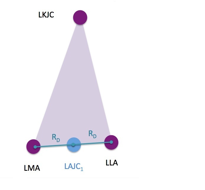
In this example, three border targets are used. So the frontal plane is simply the plane defined by the three targets.

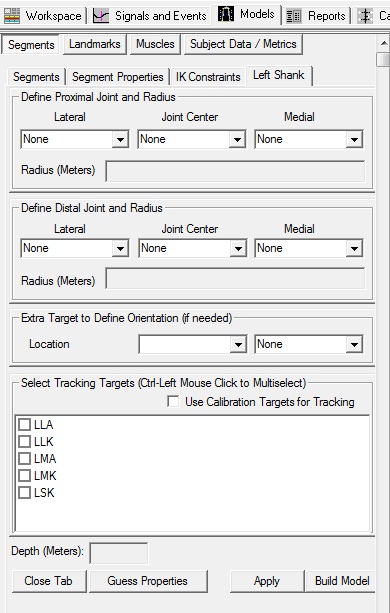
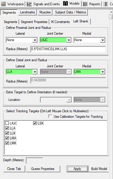
For example of Method 1, create the left shank segment with the following definition:
1. Create Left Shank Segment:


  1. In the Segments tab, select Left Shank in the Segment Name box.


  2. Click on the Create Segment button.


  3. In the Left Shank tab, enter these values:

       Define Proximal Joint and Radius
       Lateral: None     Joint: LKJC     Medial: None     
       Radius: 0.5*DISTANCE(LMK,LLK)


       Define Distal Joint and Radius
       Lateral: LLA     Joint: None     Medial: LMA     


       Select Tracking Targets:
         LLA, LLK, LMA, LMK, LSK


  4. Click on Build Model. A 3D image of a shank will appear in the 3D viewer.


  5. Click on Close Tab before proceeding.

    segmentdefinition_proxjc_distlatmed.jpg


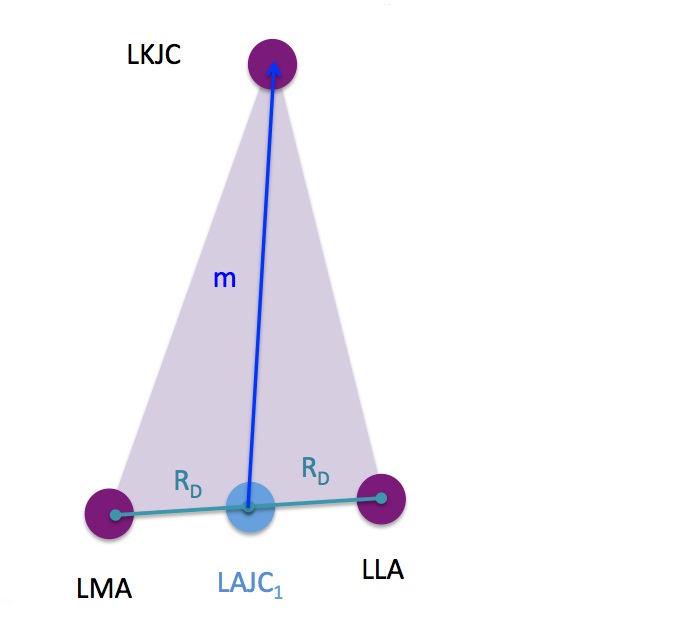
    When using a proximal joint center and the mediolateral distal targets to define the segment, the distal joint center is defined by:
    1) Define the anatomical plane (purple triangle) using the three targets
    2) Find the midpoint between the mediolateral targets
    3) The distal joint center is defined at the midpoints

    segmentdefinition_proxjc_distlatmed_ppt.jpg

    Method 2

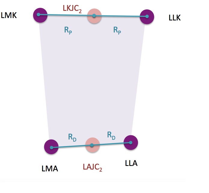
    In this example, four border targets are used. A plane is defined by three points, so when four targets are used, the frontal plane is fit between the four targets using a least square fit. The least squares fit is applied such that the sum of squares distance between the targets and the frontal plane is minimized.

    For an example of Method 2, create the left shank segment with the following definition:
    2. Create Left Shank Segment:


    1. In the Segments tab, select Left Shank in the Segment Name box.


    2. Click on the Create Segment button.


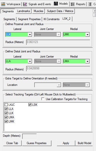
    3. In the LSK_2 tab, enter these values:

         Define Proximal Joint and Radius
         Lateral: LLK     Joint: None     Medial: LMK     


         Define Distal Joint and Radius
         Lateral: LLA     Joint: None     Medial: LMA     


         Select Tracking Targets:
           LLA, LLK, LMA, LMK, LSK


    4. Click on Build Model. A 3D image of a shank will appear in the 3D viewer.


    5. Click on Close Tab before proceeding.

      segmentdefinition_proxlatmed_distlatmed.jpg

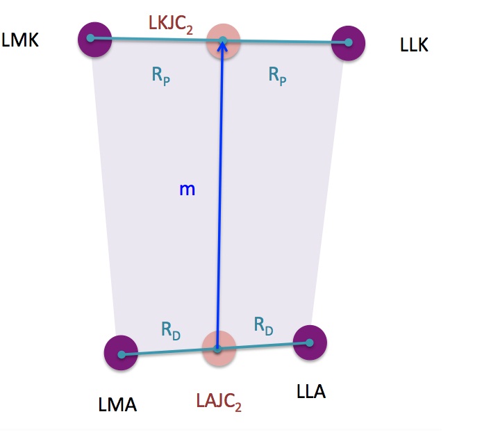
      When using a proximal joint center and the mediolateral distal targets to define the segment, the distal joint center is defined by:
      1) Define the anatomical plane (purple box) using the least squares fit to define the plane between the four targets
      2) Find the midpoint between the proximal mediolateral targets
      3) The proximal joint center is defined at the midpoint
      4) Find the midpoint between the distal mediolateral targets
      5) The distal joint center is defined at the midpoint

      segmentdefinition_proxlatmed_distlatmed_ppt.jpg

      Method 3

      In this example, three border targets are used. So the frontal plane is simply the plane defined by the three targets.

      For example of Method 3, create the left shank segment with the following definition:
      3. Create Left Shank Segment:


      1. In the Segments tab, select Left Shank in the Segment Name box.


      2. Click on the Create Segment button.


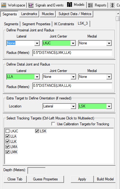
      3. In the LSK_3 tab, enter these values:

           Define Proximal Joint and Radius
           Lateral: None     Joint: LKJC     Medial: None     
           Radius: 0.5*DISTANCE(LLK,LMK)


           Define Distal Joint and Radius
           Lateral: LLA     Joint: None     Medial: None     
           Radius: 0.5*DISTANCE(LLA,LMA)


           Extra Target to Define Orientation
           Location: Lateral     LSK


           Select Tracking Targets:
             LLA, LLK, LMA, LMK, LSK


      4. Click on Build Model. A 3D image of a shank will appear in the 3D viewer.


      5. Click on Close Tab before proceeding.

        segmentdefinition_proxjc_distlat.jpg

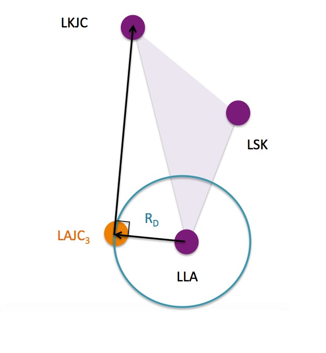
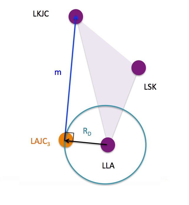
        When using a proximal joint center and the lateral distal target to define the segment:
        1) Define the anatomical plane (purple triangle) using the three targets
        2) Draw a circle with radius “Rd” around the lateral distal target
        D 3) Draw a vector from the proximal joint center that forms a right angle with the radius of the circle
        4) The joint center is at the location where the two vectors meet

        segmentdefinition_proxjc_distlat_ppt.jpg

        Method 4

        In this example, three border targets are used. So the frontal plane is simply the plane defined by the three targets.

        For example of Method 4, create the left shank segment with the following definition:
        4. Create Left Shank Segment:


        1. In the Segments tab, select Left Shank in the Segment Name box.


        2. Click on the Create Segment button.


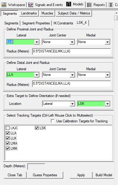
        3. In the LSK_4 tab, enter these values:

             Define Proximal Joint and Radius
             Lateral: LLK     Joint: None     Medial: None     
             Radius: 0.5*DISTANCE(LLK,LMK)


             Define Distal Joint and Radius
             Lateral: LLA     Joint: None     Medial: None     
             Radius: 0.5*DISTANCE(LLA,LMA)


             Extra Target to Define Orientation
             Location: Lateral     LSK


             Select Tracking Targets:
               LLA, LLK, LMA, LMK, LSK


        4. Click on Build Model. A 3D image of a shank will appear in the 3D viewer.


        5. Click on Close Tab before proceeding.

          segmentdefinition_proxlat_distlatmed.jpg

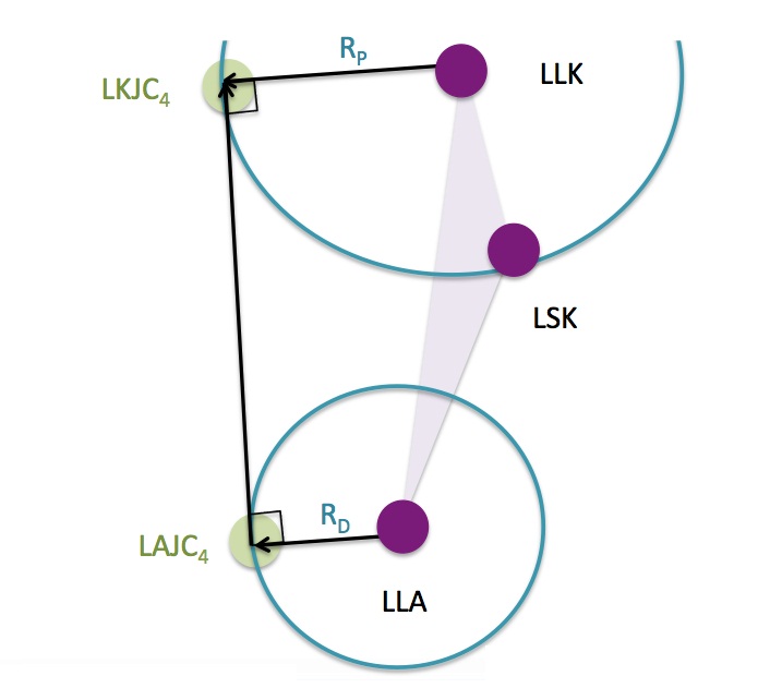
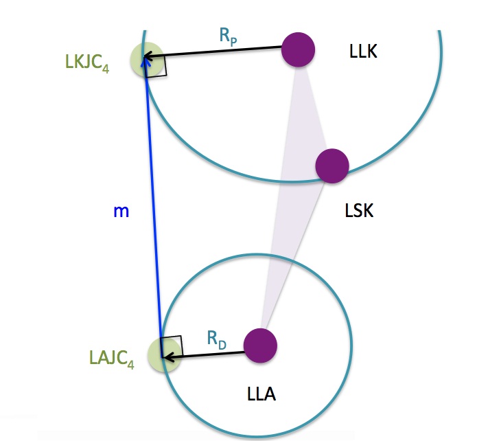
          When using a proximal joint center and the lateral distal target to define the segment:
          1) Define the anatomical plane (purple triangle) using the three targets
          1) Draw a circle with radius “Rp” around the lateral proximal target
          2) Draw a circle with radius “Rd” around the lateral distal target
          3) Drawing a vector on the “medial” side of the circle that forms a right angle with the radius of the proximal and distal circles
          4) The joint centers are at the locations where the vector meet the radii

          segmentdefinition_proxlat_distlat_ppt.jpg

          InferiorSuperior Axis Orientation

          The Inferior/Superior is aligned with the vector which is drawn from the distal joint center to the proximal joint center.

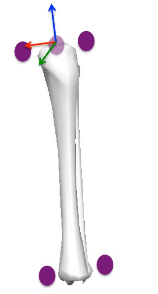
          By default, the inferior/superior axis is the Z-Axis (drawn in blue), but different labs may use a different conventions.
          For the four methods listed above, the inferior/superior axis of the segment coordinate system will be aligned with the vector “m” drawn in the images below:

          Segment Coordinate System Definition

          Visual3D will then create the segment coordinate system using the anatomical plane and joint centers defined. Explanation of the segment coordinate system is defined here. ppt_segmentcoordinatesystem.jpg
          visual3d/documentation/modeling/coordinate_system.txt · Last modified: 2024/07/17 11:45 by sgranger