CalibrateDSX: The Importance of Image Smoothing and Downsampling
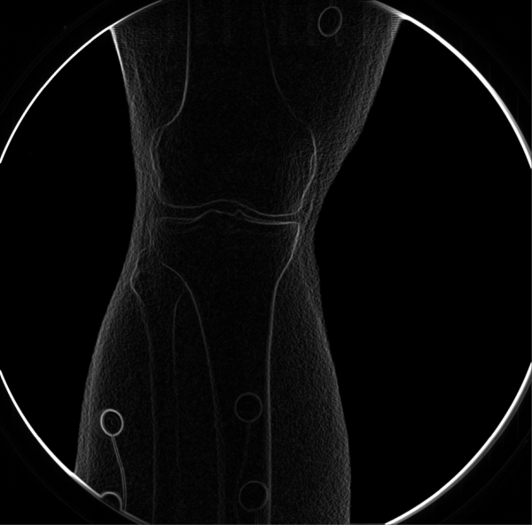
DRR-based tracking of bones in X-ray images relies heavily on the brightness of the bones' edges in the edge-detected images. Smoothing and/or resizing the images during correction increases their signal-to-noise ratio, resulting in brighter edges. Some amount of smoothing or resizing is almost always needed to maximize the trackability of the bones.
Example: Knee
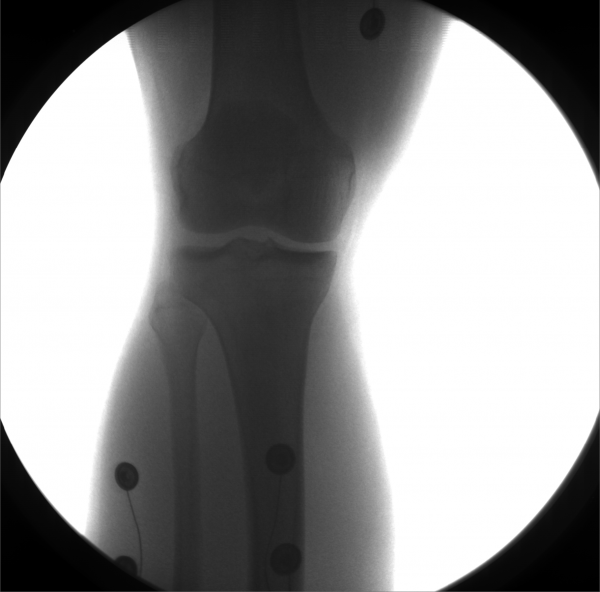
These images were captured at the Biodynamics Lab at the University of Pittsburgh. They are 14-bit grayscale with a resolution of 1824x1800 pixels.
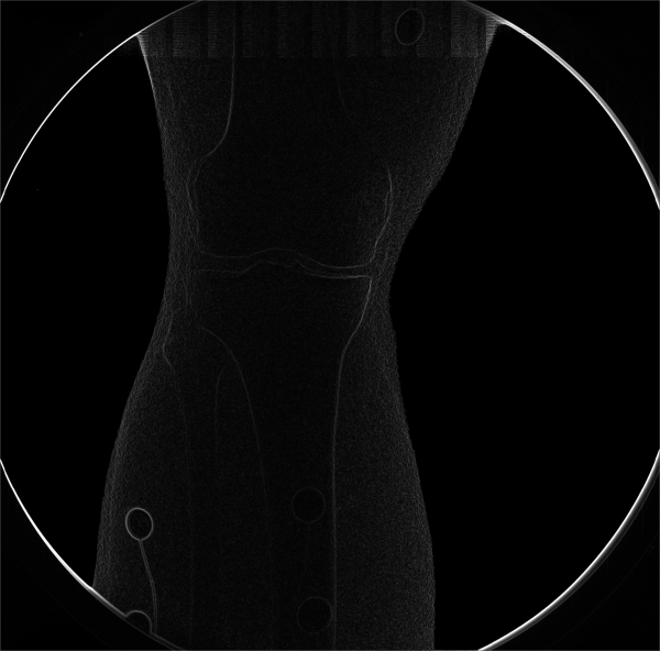
Original and edge-detected:
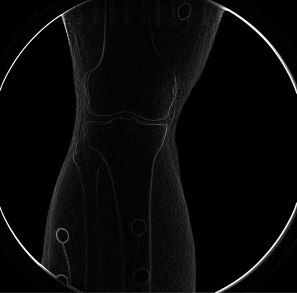
Smoothed 5x5, sigma = 2.0 (left), scaled 0.5 (right):
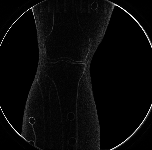
Smoothed 5x5, sigma = 2.0 and scaled 0.5:
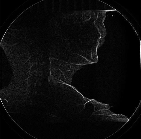
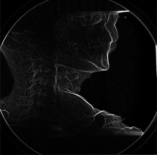
Example: Cervical Spine
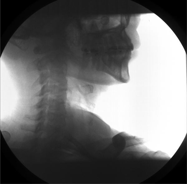
These images were captured at the Biodynamics Lab at the University of Pittsburgh. They are 14-bit grayscale with a resolution of 1824x1800 pixels.
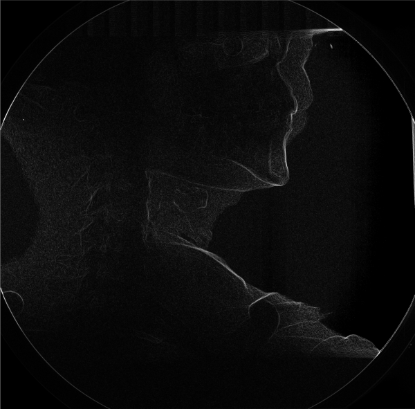
Original and edge-detected:
Smoothed 5x5, sigma = 2.0 (left), scaled 0.5 (right):
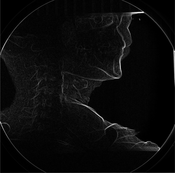
Smoothed 5x5, sigma = 2.0 and scaled 0.5:
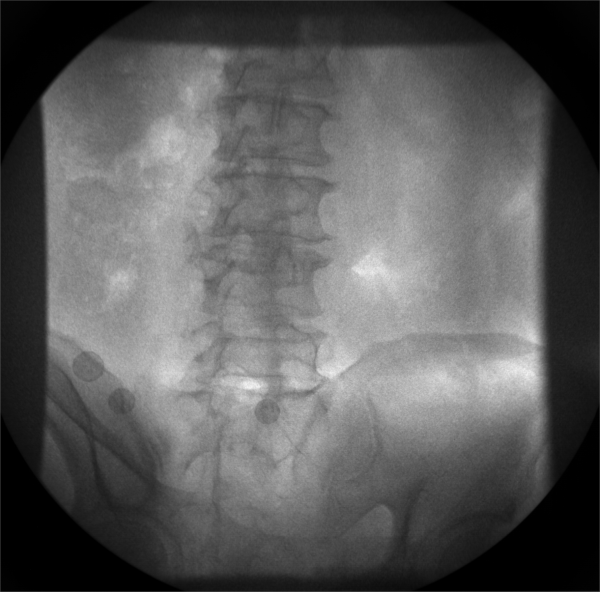
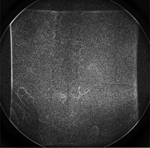
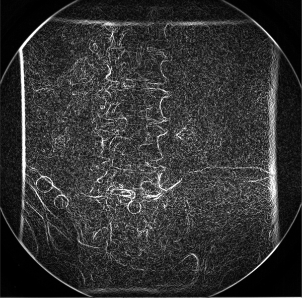
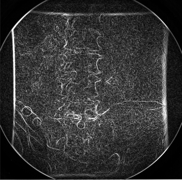
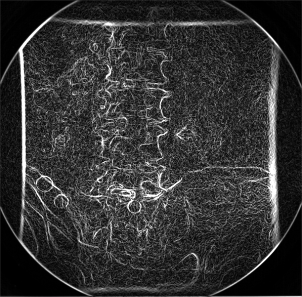
Example: LumbarSpine
These images were captured at the Biodynamics Lab at the University of Pittsburgh. They are 14-bit grayscale with a resolution of 1824x1800 pixels.
Original and edge-detected:
Smoothed 7x7, sigma = 4.0 (left), scaled 0.33 (right):
Smoothed 7x7, sigma = 4.0 and scaled 0.33:
